
Anatomy and Physiology Course Notes
Comprehensive Guide for BSN 1st Year Students - 1st Semester
Unit 1 – Introduction to Anatomy and Physiology
Anatomy is the study of body structure; it examines how body parts are organized and their relationships to one another.
Physiology studies the functions of these structures and how they work individually and together to maintain life.
Normal Anatomical Position and Significance
The standard anatomical position provides a universal reference for describing body structures:
- Body standing upright
- Feet parallel and flat on the floor
- Arms at the sides
- Palms facing forward with thumbs pointing away from the body

Standard Anatomical Position (Source: Wikimedia Commons)
This consistent reference point prevents confusion when locating structures or describing injuries. When the body is in any other position, directional terms still refer to this standard position.
Body Planes and Directional Terminology

Major Body Planes (Source: Wikimedia Commons)
Body planes are imaginary flat surfaces that slice through the body:
- Sagittal plane - divides the body into left and right portions
- Midsagittal plane - exactly midline, creating equal left and right halves
- Frontal (coronal) plane - divides the body into anterior (front) and posterior (back) sections
- Transverse (horizontal) plane - divides the body into superior (upper) and inferior (lower) parts
Directional Terms
These terms describe the location of body structures relative to each other:
| Term | Definition | Example |
|---|---|---|
| Anterior/Ventral | Toward the front of the body | The sternum is anterior to the spine |
| Posterior/Dorsal | Toward the back of the body | The spine is posterior to the sternum |
| Superior | Above or closer to the head | The head is superior to the neck |
| Inferior | Below or closer to the feet | The feet are inferior to the knees |
| Medial | Toward the midline of the body | The nose is medial to the eyes |
| Lateral | Away from the midline of the body | The ears are lateral to the nose |
| Proximal | Closer to the point of attachment | The elbow is proximal to the wrist |
| Distal | Farther from the point of attachment | The fingers are distal to the wrist |
Body Cavities
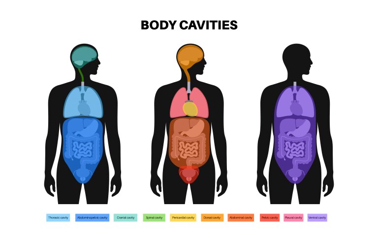
Major Body Cavities (Source: Wikimedia Commons)
The body contains several major cavities that protect organs and allow them to expand and contract:
- Dorsal cavity - includes the cranial cavity (brain) and vertebral canal (spinal cord)
- Ventral cavity - divided into:
- Thoracic cavity (pleural cavities, mediastinum, pericardial cavity)
- Abdominopelvic cavity (abdominal and pelvic cavities)
The diaphragm separates the thoracic and abdominal cavities.
Abdominopelvic Quadrants and Regions

Abdominopelvic Quadrants and Regions (Source: Wikimedia Commons)
Clinicians divide the abdomen for diagnostic purposes:
- Four Quadrants - created by vertical and horizontal lines through the umbilicus:
- Right Upper Quadrant (RUQ) - liver, gallbladder
- Left Upper Quadrant (LUQ) - stomach, spleen
- Right Lower Quadrant (RLQ) - appendix, right ovary
- Left Lower Quadrant (LLQ) - left ovary, sigmoid colon
- Nine Regions - more detailed division using two horizontal and two vertical lines:
- Right hypochondriac, Epigastric, Left hypochondriac
- Right lumbar, Umbilical, Left lumbar
- Right iliac, Hypogastric, Left iliac
Unit 2 – Homeostatic Mechanisms and Clinical Significance
Homeostasis refers to the body's ability to maintain a stable internal environment despite changes in the external environment.
Homeostasis is a dynamic process involving continuous monitoring and adjustment of physiological variables. For example, body temperature remains around 37°C through mechanisms like sweating or shivering.
Components of Feedback Systems
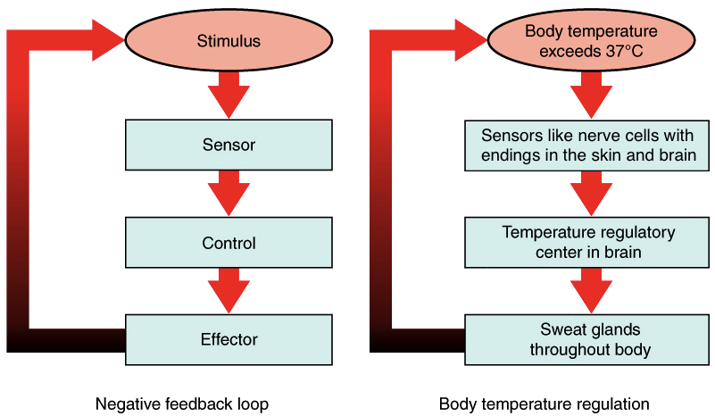
Negative Feedback Loop Mechanism (Source: Wikimedia Commons)
Homeostatic control operates through feedback loops with three components:
- Sensor (Receptor) - detects changes in physiological values (e.g., thermoreceptors detecting cold)
- Control Center - compares sensor input to set point and sends instructions (often the brain or endocrine gland)
- Effector - produces a response that returns the value toward the set point (e.g., muscles shivering to generate heat)
Types of Feedback Loops
| Type | Mechanism | Examples |
|---|---|---|
| Negative Feedback | Reverses deviations from set point; promotes stability | Body temperature regulation, Blood glucose control, Blood pressure regulation |
| Positive Feedback | Amplifies initial change until endpoint is reached | Childbirth (oxytocin release), Blood clotting, Action potential generation |
Clinical Significance
Understanding feedback systems helps nurses anticipate patient responses:
- Failure of negative feedback mechanisms can cause hypertension or hyperthermia
- Positive feedback can be beneficial (e.g., platelet aggregation in clotting) but harmful if uncontrolled (e.g., cytokine storms in sepsis)
- Many medications work by modifying feedback systems (e.g., insulin for diabetes, antihypertensives for high blood pressure)
Unit 3 – Musculoskeletal System
Divisions of the Skeleton

Human Skeleton - Axial and Appendicular Divisions (Source: Wikimedia Commons)
| Division | Components | Function | Number of Bones |
|---|---|---|---|
| Axial Skeleton | Skull, vertebral column, thoracic cage, hyoid bone, ear ossicles | Supports and protects brain, spinal cord, heart, and lungs; provides attachment sites for muscles | 80 bones |
| Appendicular Skeleton | Upper and lower limbs, pectoral and pelvic girdles | Facilitates movement; lower limbs adapted for weight-bearing, upper limbs for mobility and dexterity | 126 bones |
Classification of Joints
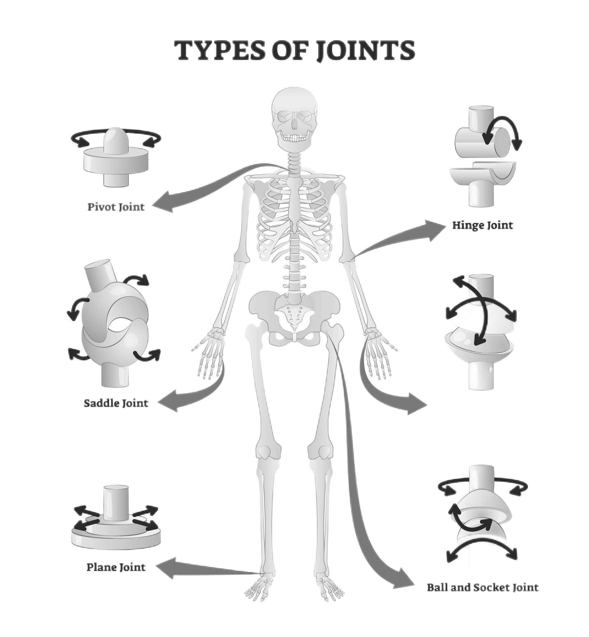
Types of Synovial Joints (Source: Wikimedia Commons)
Joints are classified both structurally and functionally:
Structural Classification
- Fibrous joints - bones joined by dense connective tissue; mostly immovable (e.g., skull sutures)
- Cartilaginous joints - bones connected by cartilage; allow limited movement (e.g., pubic symphysis, intervertebral discs)
- Synovial joints - contain a fluid-filled synovial cavity; permit free movement (e.g., shoulder, knee, hip)
Functional Classification
- Synarthrosis - immovable joints (e.g., skull sutures)
- Amphiarthrosis - slightly movable joints (e.g., intervertebral discs)
- Diarthrosis - freely movable joints; all synovial joints fall into this category
Muscle Types and Characteristics

Three Types of Muscle Tissue (Source: Wikimedia Commons)
| Muscle Type | Structure | Control | Function | Location |
|---|---|---|---|---|
| Skeletal Muscle | Long, cylindrical, multinucleated, striated | Voluntary | Movement, posture, heat production | Attached to bones |
| Cardiac Muscle | Branched, striated, single nucleus, intercalated discs | Involuntary | Pumps blood through circulatory system | Heart |
| Smooth Muscle | Spindle-shaped, non-striated, single nucleus | Involuntary | Moves substances through internal passageways | Walls of hollow organs, blood vessels |
Unit 4 – Integumentary System
Skin Layers and Functions

Layers of the Skin (Source: Wikimedia Commons)
The skin has two main layers:
- Epidermis - outer layer of keratinized stratified squamous epithelium
- Dermis - inner layer of dense irregular connective tissue containing blood vessels, nerves, and accessory structures
Deep to the dermis is the hypodermis (subcutaneous layer) consisting of loose connective and adipose tissue.
Epidermal Layers (from deep to superficial):
- Stratum basale - deepest layer with active cell division
- Stratum spinosum - several layers of keratinocytes
- Stratum granulosum - cells accumulate keratin and begin to degenerate
- Stratum lucidum - clear layer found only in thick skin (palms, soles)
- Stratum corneum - outermost layer of dead, keratinized cells that are continuously shed
Accessory Structures

Hair Follicle Structure (Source: Wikimedia Commons)
- Hair - keratinous filament originating from hair follicles; provides protection, sensory input, and thermoregulation
- Nails - plates of densely packed dead keratinocytes; protect distal phalanges and assist with manipulation
- Sweat glands -
- Eccrine glands - produce watery sweat for thermoregulation
- Apocrine glands - secrete thicker fluid rich in organic molecules; associated with hair follicles
- Sebaceous glands - produce sebum (oil) to lubricate skin and hair
Effects of Aging on the Integumentary System
- Epidermis thins and stem cell division slows, making skin more prone to injury
- Collagen in dermis decreases, leading to wrinkles and loss of elasticity
- Reduced glandular activity results in dry skin
- Melanocyte activity declines, causing hair to turn gray
- Wound healing slows due to decreased blood supply and cell proliferation
Unit 5 – Role of Blood in Human Physiology
Functions of Blood

Composition of Blood (Source: Wikimedia Commons)
Blood is a specialized connective tissue with multiple vital functions:
- Transportation - delivers oxygen, nutrients, hormones; removes wastes like carbon dioxide and urea
- Defense - leukocytes protect from pathogens; platelets and plasma proteins form clots
- Homeostasis - distributes heat, regulates pH through buffers, maintains fluid balance
Composition of Blood
| Component | Percentage | Composition | Function |
|---|---|---|---|
| Plasma | 55% | Water (92%), proteins (7%), other solutes (1%) | Transport medium, clotting, immunity, pH balance |
| Formed Elements | 45% | Erythrocytes, leukocytes, platelets | Gas transport, immunity, clotting |
Major Plasma Proteins:
- Albumin - maintains osmotic pressure, transports substances
- Globulins - include antibodies (immunoglobulins) and transport proteins
- Fibrinogen - essential for blood clotting
Structure and Function of Cellular Components

Blood Cells: Erythrocytes, Leukocytes, and Platelets (Source: Wikimedia Commons)
- Erythrocytes (RBCs) - biconcave discs lacking nuclei; contain hemoglobin for oxygen transport
- Leukocytes (WBCs) - defend against pathogens; types include neutrophils, lymphocytes, monocytes, eosinophils, basophils
- Platelets - cell fragments derived from megakaryocytes; essential for hemostasis
Clinical Significance of Blood Proteins
- Low albumin levels - can lead to edema due to decreased osmotic pressure; seen in liver disease or malnutrition
- Elevated globulins - may indicate chronic inflammation or immune disorders
- Reduced fibrinogen - impairs clotting and increases bleeding risk
Significance of Blood Groups

ABO Blood Group System (Source: Wikimedia Commons)
Blood groups are determined by antigens on erythrocyte membranes:
ABO Blood Group System:
- Type A - has A antigens and anti-B antibodies
- Type B - has B antigens and anti-A antibodies
- Type AB - has both A and B antigens, no antibodies (universal recipient)
- Type O - has no A or B antigens, both anti-A and anti-B antibodies (universal donor)
Rh Blood Group System:
- Presence of Rh (D) antigen denotes Rh positivity
- Absence denotes Rh negativity
- Rh incompatibility between an Rh− mother and Rh+ fetus can cause hemolytic disease of the newborn
Clinical Alert: Transfusion Reactions
Transfusion of incompatible blood can lead to agglutination (clumping) and hemolysis (rupture) of RBCs, causing:
- Blockage of blood vessels
- Kidney failure due to hemoglobin release
- Fever, chills, nausea, potentially fatal shock
Always verify blood type compatibility before transfusion!
Conclusion
These comprehensive course notes integrate fundamental anatomical and physiological principles with clinical examples relevant to nursing practice. Understanding how body structures relate to their functions, how homeostatic mechanisms maintain internal constancy, and how organ systems interact provides a foundation for patient assessment and intervention.
Regular review of these concepts, along with practical demonstrations using anatomical models, will prepare BSN students for both examinations and clinical application. Remember that anatomy and physiology form the foundation of nursing practice - a thorough understanding enables better patient care, accurate assessment, and appropriate intervention.